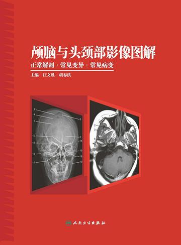
1. Taking into account the portability of the book and the needs of clinical specialists, this book has several volumes. This volume is the cranium and head and neck volume;
2. The content is organized according to the main line of parts, and the division and integration between different equipment are combined. For example, X-ray anatomy is written separately and includes angiography; CT and MRI anatomy are written together. The content of the sagittal and coronal planes is mainly MRI, and is connected with clinical practice. There are trade-offs, and it is not exhaustive;
3. Combine a normal picture with a diagrammatic picture, and use reconstructed pictures or schematic diagrams to indicate the tangent lines of each picture. When typesetting, compare the pictures on one side with the text on the other, and the text should be concise, including picture descriptions and impressions, anatomical points (structures and characteristics that can be shown in the pictures), important measurement data, normal variations, etc.;
4. Unlabeled unillustrated pictures are arranged next to the illustrated pictures to facilitate readers' understanding;
5. After the introduction of each part, some analysis of the most common lesions is appropriately written, so that readers can learn to transition from normal to looking at lesions, making this book the basis and bridge for readers to learn to look at disease films, which is the biggest feature of this book;
6. The selection of common cases is based on important structures, common diseases in locations, diseases that are discovered or can be diagnosed based on changes in anatomical structures, etc.